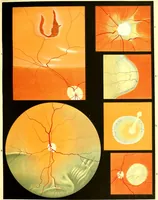
Atlas of ophthalmoscopy : Representing the normal and pathological conditions of the fundus oculi as seen with the ophthalmoscope. Composed of 12 chromolithographic plates, containing 59 figures, drawn from nature and accompanied by an explanatory text / by Dr. R. Liebreich, the text translated by H. Rosborough Swanzy.
A medical atlas of ophthalmoscopy by Richard Liebreich, drawn from life and issued in English translation by H. Rosborough Swanzy in London in 1870 for eye surgeons and students. Twelve chromolithographic plates show the fundus of the eye as circular and square fields of flat orange and vermilion, crossed by branching red vessels and pale yellow discs, hemorrhage blots and white exudate patches, offering designers ready-made radial vessel diagrams and high-contrast warm colour fields.