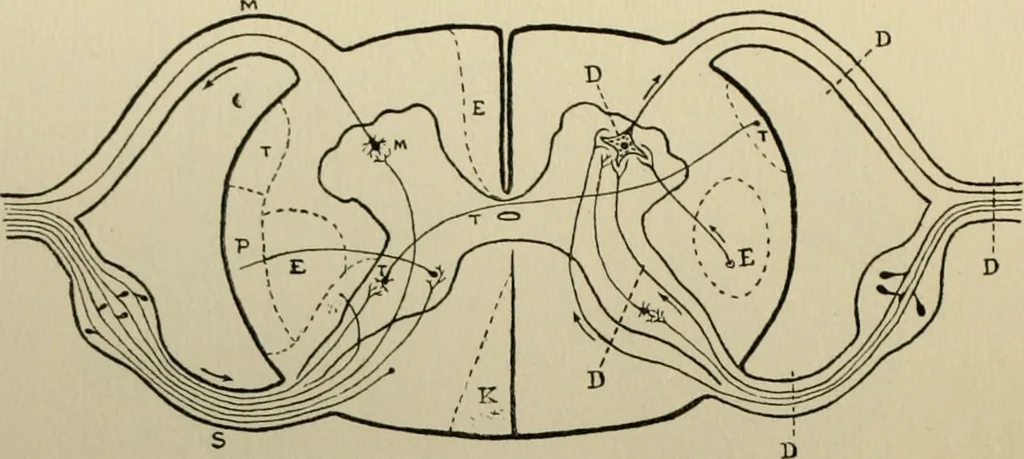
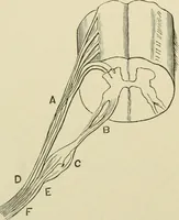
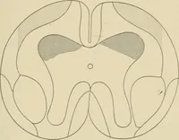
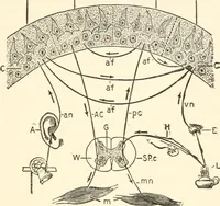
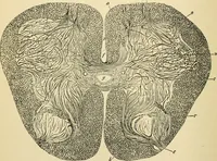
uft at tin- lirn- of junrtion with the medulla, showing the motor decussation and thetopography of the uppermost level of the cord. Bh L208 24fi … Fig. 24 DIAGRAMMATIC SECTION OF THE SPINAL CORD TO ILLUSTRATEITS PHYSIOLOGY Left side shows situation of lesions causingdisorders of motion and sensation. Right side shows situation of lesions causingdisorders of reflex activity. Destructive lesions at M or E cause diminution, slight irritative…
Atlases · Nervous System Diseases
Internet Archive · Medical Heritage Library · Public Domain Mark 1.0. Free to use for anything.