Fractures and dislocations, diagnosis and treatment (1915)
Preston, Miller Edwin, 1879- [from old catalog], Stover, H. G. [from old catalog] · St. Louis, C. V. Mosby company · leaf 562
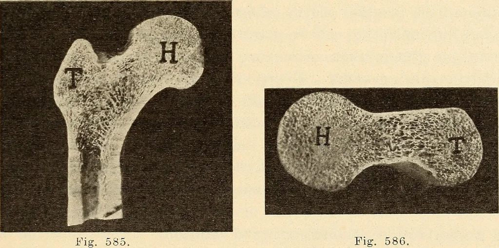
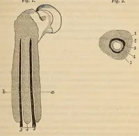
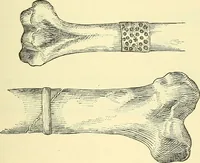
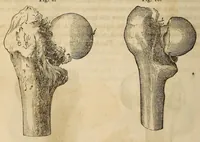
centers of ossification; one for the head, one for thegreater, and one for the lesser trochanter. They all join thediaphysis at about the eighteenth year. Epiphyseal separations,however, are extremely rare in the upper end of this bone, exceptas a chronic condition in children, which is known as… … Fig. 586. Figs. 585 and 586.—Coronal and nearly horizontal sections of the upper end ofthe femur showing the relative distribution of cancellous and compact tissue. H., headof femur. T., trochanter major. (Preston—Surg., Gynecol, and Obstet., Feb., 1914.) of the clinical conditions occurring in…
Fractures · Dislocations · X-rays
Internet Archive · Library of Congress · Public Domain Mark 1.0. Free to use for anything.