Oral Roentgenology : a Roentgen study of the anatomy and pathology of the oral cavity (1917)
Thoma, Kurt H. (Kurt Hermann), 1883- · Boston, Mass. : Ritter & Company · leaf 100
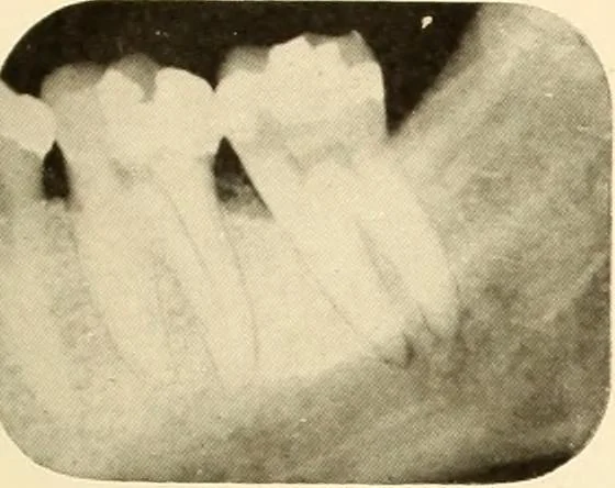
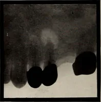
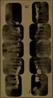
pain felt at angle of jaw. Examination of mouth showed well-cared-forteeth with large fillings. Roentgen Examination: Shows a radiolucent area of well defined character on the roots of thelower first molar. A dark area under distal part of filling indicates decay. The conclusion from thesefindings…