Oral Roentgenology : a Roentgen study of the anatomy and pathology of the oral cavity (1917)
Thoma, Kurt H. (Kurt Hermann), 1883- · Boston, Mass. : Ritter & Company · leaf 104
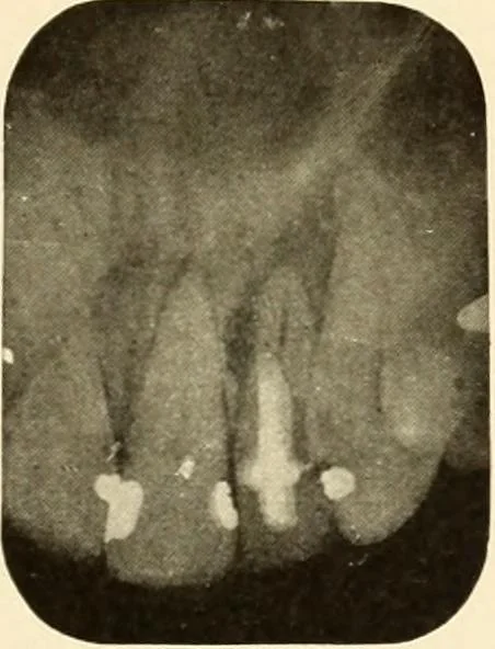
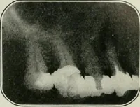
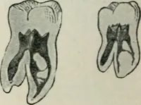
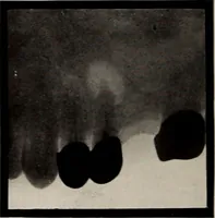
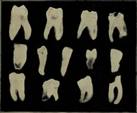
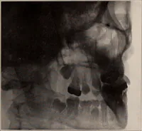
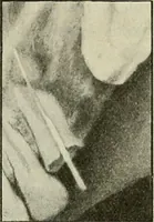
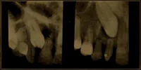
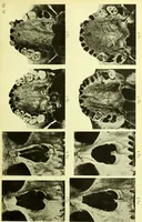
Examination: Shows lateral granuloma, caused by perfor-ation at the side of the root. Figures 116-118. History: None of these three cases caused any discomfort and theteeth had been devitalized for varying lengths of time. Roentgen Examination: Shows incomplete root canal filhngs. Theradiolucent… … Figure ^*1&}