Oral Roentgenology : a Roentgen study of the anatomy and pathology of the oral cavity (1917)
Thoma, Kurt H. (Kurt Hermann), 1883- · Boston, Mass. : Ritter & Company · leaf 138
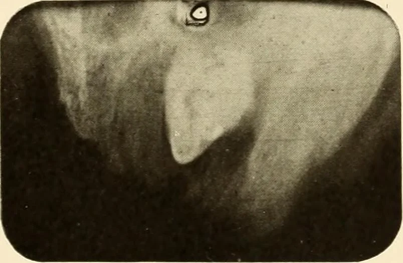
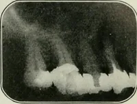
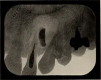
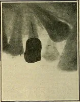
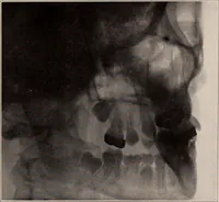
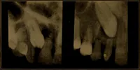
Figure … Figure 134 ORAL ROENTGENOLOGY Roentgenology of Dentigerous Cysts Figures 187 and 188. Patient: Mrs. H. H. P. History: Upper cuspid has never erupted. The lateral incisor hasbeen sore at times. Roentgen Examination: Shows a radiopaque object which seems tobe the missing tooth. Its cusp is in contact…