Oral Roentgenology : a Roentgen study of the anatomy and pathology of the oral cavity (1917)
Thoma, Kurt H. (Kurt Hermann), 1883- · Boston, Mass. : Ritter & Company · leaf 142
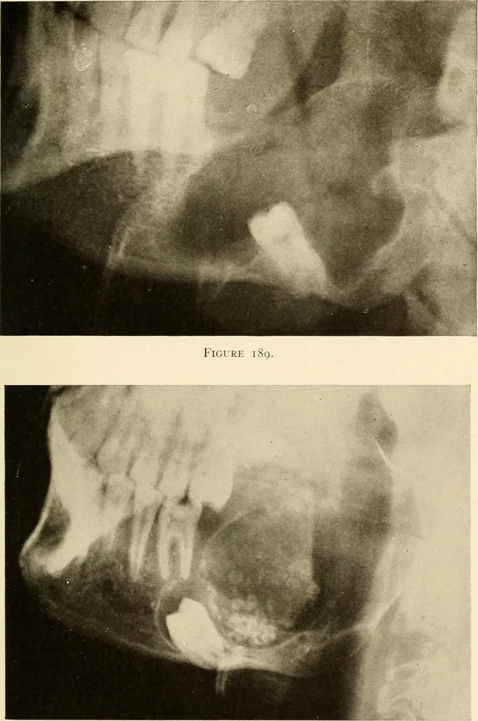
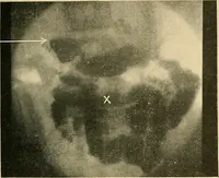
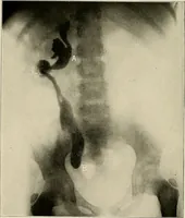
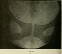
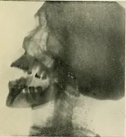
bone. The cyst, itself, was thin-walled in its upper part and nearly | inchthick in its lower part. In the course of the separation it was ruptured, and strawcolored fluid escaped. This was removed, leaving a smooth-walled cavity in the bone. Result of Operation: A Roentgenogram taken two months… … Figure 190. 138 ORAL ROENTGENOLOGY Roentgenology of Simple and Multilocular Periodontal Cysts Figure 191. Patient: Mr. J. T. G. History: Patient complained of tender place on outside of face which he noticedespecially when shaving. Roentgen Examination: Shows a large radiolucent area with…