Oral Roentgenology : a Roentgen study of the anatomy and pathology of the oral cavity (1917)
Thoma, Kurt H. (Kurt Hermann), 1883- · Boston, Mass. : Ritter & Company · leaf 148
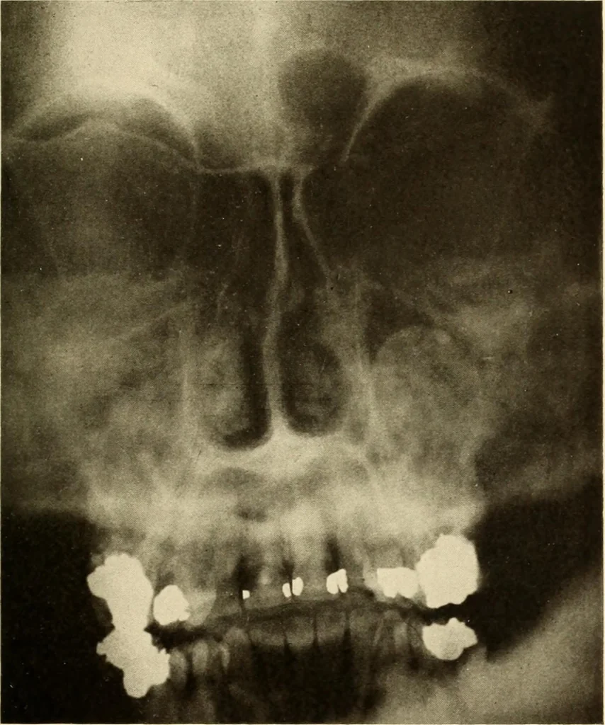
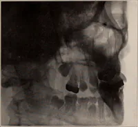
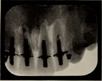
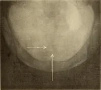
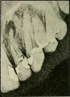
trum, on the right side a radiopaque structure of definite shapewith a well defined outline. The upper part of the antrum on this sideis normal. Operative Findings: The right maxillary sinus was found to be filledby a periodontal cyst, originating from abscessed teeth in the upper jaw.After the… … Figure 195 144 ORAL ROENTGENOLOGY Roentgenology of Multilocular Cysts Figures 196 and 197. Patient: Mr. R. W., age 19 years. Courtesy of Dr. Samuel Mixterand Dr. G. D. Cutler. History: When one year of age was hit hard on jaw with pump handleand was said to have fractured jaw at that time. Since…