Oral Roentgenology : a Roentgen study of the anatomy and pathology of the oral cavity (1917)
Thoma, Kurt H. (Kurt Hermann), 1883- · Boston, Mass. : Ritter & Company · leaf 150
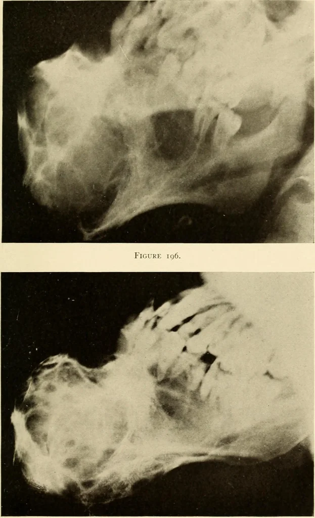
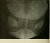
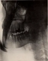
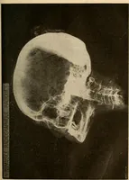
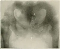
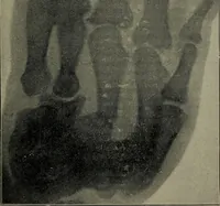
man. Large, hard, irregulartumor found on lower jaw, mostly on right side, so firmly attached asto be continuous with the jaw bone. Skin of tumor not adherent, abnor-mal nor tender except at right corner of mouth, which was the site of theabscess. Roentgen Examination: Roentgen plate by courtesy of… … Figure 197. 146 ORAL ROENTGENOLOGY Roentgenology of Carcinomata of the Jaws Figure 198. Patient: Courtesy of Dr. L. B. Morison. History: The tumor started as an epithehoma of the Up. Themetastatic processes of the carcinoma affected the jaw, as seen in theRoentgen picture. Roentgen Examination:…