Oral Roentgenology : a Roentgen study of the anatomy and pathology of the oral cavity (1917)
Thoma, Kurt H. (Kurt Hermann), 1883- · Boston, Mass. : Ritter & Company · leaf 160
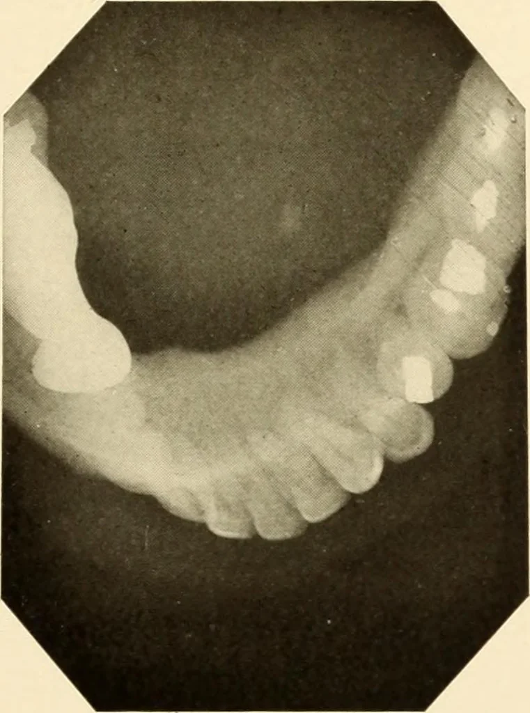
n sizeand was usually largest before meal times. Roentgen Examination: Shows a radiopaque object on the right side of the floorof the mouth opposite the first molar. Operative Findings: An incision was made in the mucous membrane of the floorof the mouth to expose the submaxillary duct and… … Figure 21 156 ORAL ROENTGENOLOGY Roentgenology of Salivary Calculi Figures 212 and 213. Patient: Mrs. H. E. M., courtesy of Dr. John T. Bottomley. History: Patient had tumor-like swelling on cheek and felt a hardsubstance under the skin, but no pain. Roentgen Examination: Plates by courtesy of Dr.…