Oral Roentgenology : a Roentgen study of the anatomy and pathology of the oral cavity (1917)
Thoma, Kurt H. (Kurt Hermann), 1883- · Boston, Mass. : Ritter & Company · leaf 172
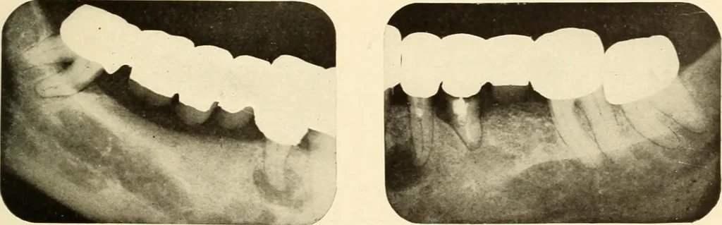
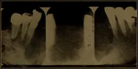
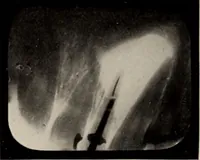
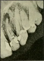
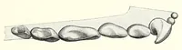
Figure 223. Figure 224. … Figure 22= Figure 226. Figure 227. 168 ORAL ROENTGENOLOGY Roentgenograms Showing Broken Root Canal Instruments Figure 228.The radiopaque object in the root canal of the upper first bicuspid was found to be a piece of a broach. Figure 229. The radiopaque object in the distal root canal of the first…