Oral Roentgenology : a Roentgen study of the anatomy and pathology of the oral cavity (1917)
Thoma, Kurt H. (Kurt Hermann), 1883- · Boston, Mass. : Ritter & Company · leaf 174
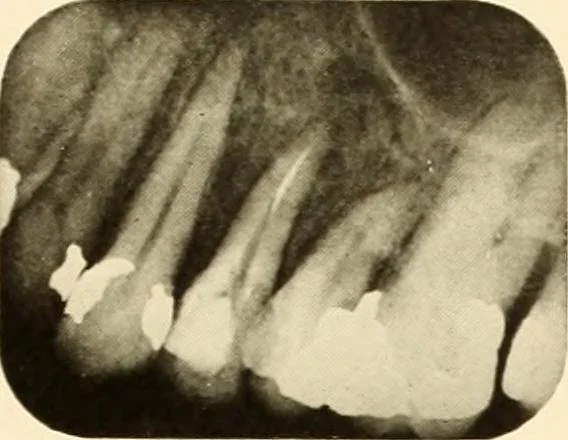
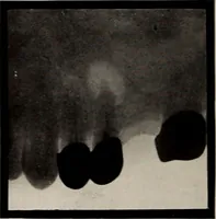
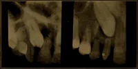
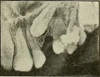
a small radiopaque objectwhich appears much lighter than the root canal filling. Roentgenograms Showing Root Perforations Figure 231. The cuspid shows the root canal filling in the proper place, but thepost of the porcelain crown protrudes on the side of the crown. Notethe radiolucent area… … Figure 228.