Oral Roentgenology : a Roentgen study of the anatomy and pathology of the oral cavity (1917)
Thoma, Kurt H. (Kurt Hermann), 1883- · Boston, Mass. : Ritter & Company · leaf 178
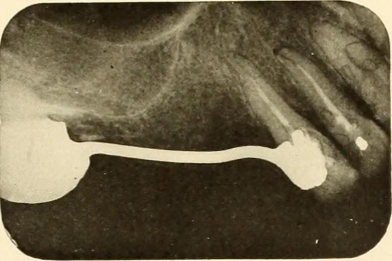
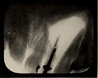
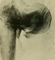
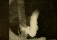
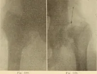
pper central incisor.Note the accessory canal and the excess of gutta percha beyond bothapical foramina. Figure 247 shows the root canal filling of an upper lateral incisor. Figure 248 shows the corkscrew variety of root canal filling in a lowerbicuspid. Figure 249 shows the filling replaced with a… … Figure 241.