Oral Roentgenology : a Roentgen study of the anatomy and pathology of the oral cavity (1917)
Thoma, Kurt H. (Kurt Hermann), 1883- · Boston, Mass. : Ritter & Company · leaf 188
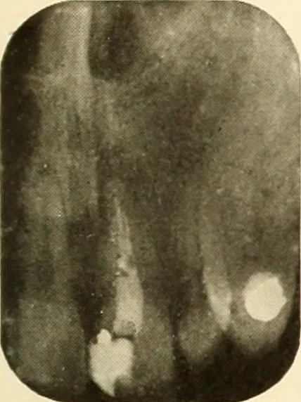
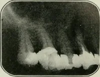
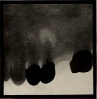
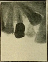
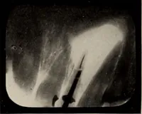
the bony wound entirely filled in, after fourteenmonths. Figures 282-286. Figure 282 shows a wire in the canal indicating the extent to whichit has been cleaned out. Figure 283 shows the canal after the filling has been put in. Figure 284 shows the crown cemented into place. Figure 285 shows the…