Oral Roentgenology : a Roentgen study of the anatomy and pathology of the oral cavity (1917)
Thoma, Kurt H. (Kurt Hermann), 1883- · Boston, Mass. : Ritter & Company · leaf 192
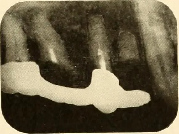
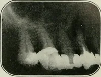
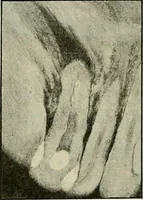
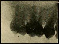
ling on gum.Roentgen Examination: Shows two infected roots under the bridge. Figures 294-296. Patient: Mr. R. S. C. History: Patient was hit with a hockey stick, fracturing the centralincisor (see Figure 294), and two fragments of the tooth were removed(see Figure 295). By means of the Roentgen… … Figure 202. Figure 293. 2^ Figure 294.