Oral Roentgenology : a Roentgen study of the anatomy and pathology of the oral cavity (1917)
Thoma, Kurt H. (Kurt Hermann), 1883- · Boston, Mass. : Ritter & Company · leaf 22
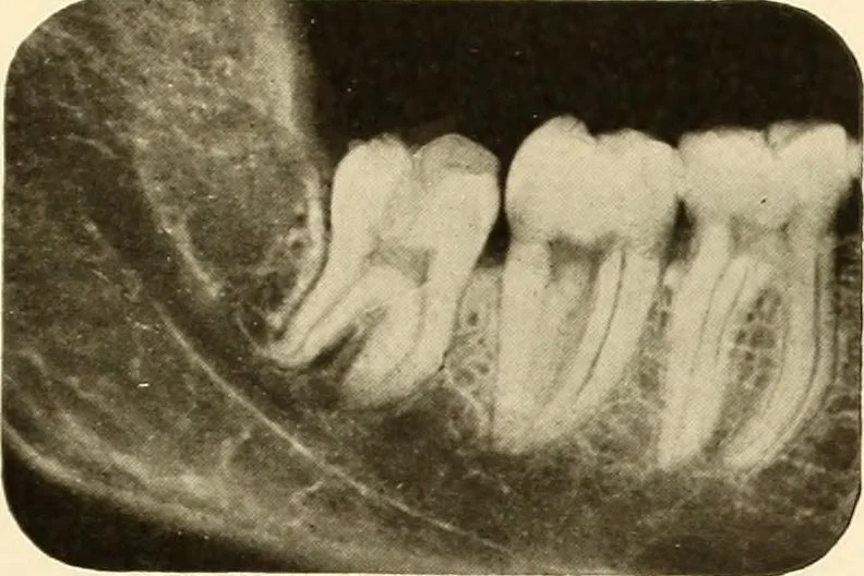
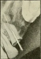
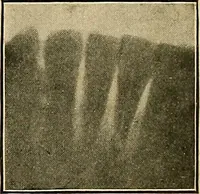
nd molar, like the others, is a perfectly healthy tooth. Thecrown, being covered by enamel, is radiopaque and shows hght in thepicture. Roentgenogram Showing a Radiolucent Area Figure 2. A cavity on the buccal side of the second molar increases the radi-ability and shows darker than the rest of the… … Figure i.