Oral Roentgenology : a Roentgen study of the anatomy and pathology of the oral cavity (1917)
Thoma, Kurt H. (Kurt Hermann), 1883- · Boston, Mass. : Ritter & Company · leaf 27
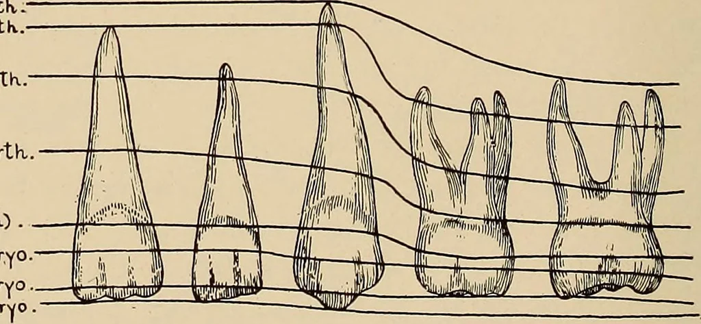
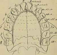
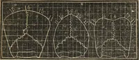
t so. Calcification of the temporary teeth is completed at the endof the second year when the roots are fully formed and the apical foraminaestablished. The diagram shown in Figure 7 gives an idea of how far thetemporary teeth have become calcified at a given age. Eruption of the Temporary Teeth.… … 40tKweekCb-irth.).30tKweek embTyo.IQtKweek embryo..I7th.week emb Progress of Calcification of the Temporary Teeth at different Periocis. ROEXTGEXOGRAPHIC STUDY OF THE XORMAL ORAL TISSUES 23 of ossification appear and these soon unite so that the contour of theprimitive jaw is estabHshed at the end…
![An illustration from [Herbier de la France, ou collection complette des plantes indigènes de ce royaume] / [Pierre Bulliard]. (1780)](https://img.insideoutlibrary.com/img/200/wellcome-wellcome_b30449248-91-1.webp)