Oral Roentgenology : a Roentgen study of the anatomy and pathology of the oral cavity (1917)
Thoma, Kurt H. (Kurt Hermann), 1883- · Boston, Mass. : Ritter & Company · leaf 34
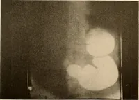
bably as a protective measure. In old age, and some-times earlier, on account of conditions such as those just mentioned, weoften find the canals of minute size and even obliterated entirely (seeFigure 32). Roentgenology of Tooth Development Figure 10. Specimen: Fetus, six months old. Roentgen… … Figure 10. 30 ORAL ROENTGENOLOGY Roentgenology of Tooth Development Figure ii. Patient: A. H., girl three years old. Roentgen Examination: All the temporary teeth have erupted.The calcification of the permanent cuspid and first molar has reachedabout two-thirds of the crowns. The tip of the first…