Oral Roentgenology : a Roentgen study of the anatomy and pathology of the oral cavity (1917)
Thoma, Kurt H. (Kurt Hermann), 1883- · Boston, Mass. : Ritter & Company · leaf 36
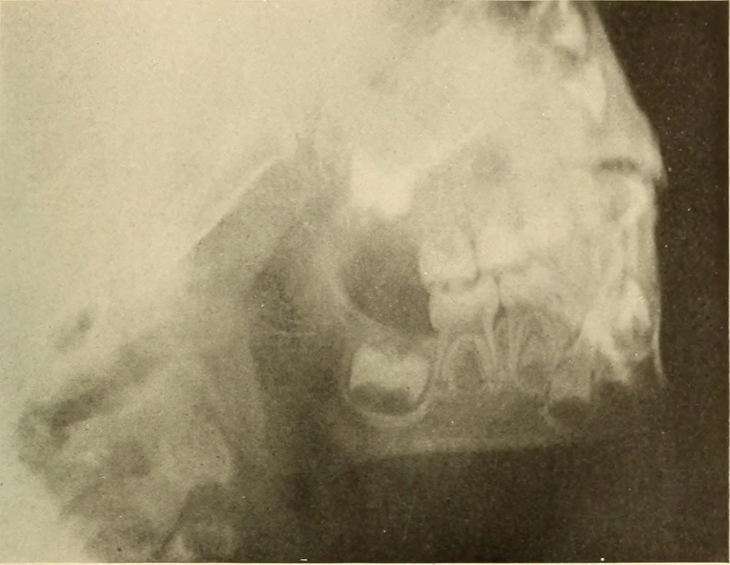
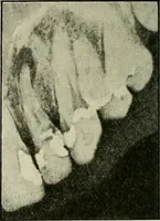
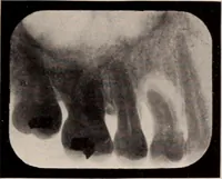
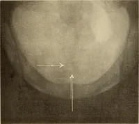
and first molar has reachedabout two-thirds of the crowns. The tip of the first bicuspid is seenbetween the roots of the first temporary molar in the lower jaw. Figure 12. Patient: H. S., girl four years old. Roentgen Examination: Here we find further progress in toothdevelopment. The roots of the… … Figure ii.