Oral Roentgenology : a Roentgen study of the anatomy and pathology of the oral cavity (1917)
Thoma, Kurt H. (Kurt Hermann), 1883- · Boston, Mass. : Ritter & Company · leaf 36
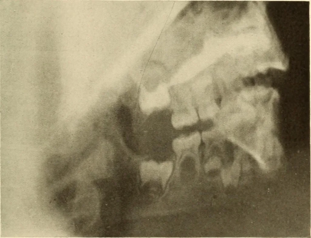
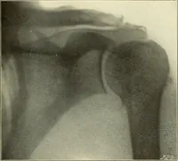
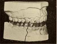
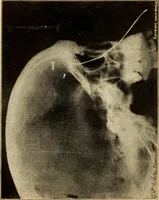
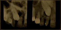
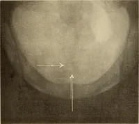
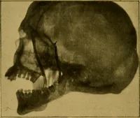
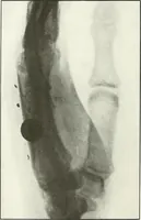
Figure ii. … Figure 12. 32 ORAL ROENTGENOLOGY Roentgenology of Tooth Development Figure 13. Patient: E. L., boy five years old. Roentgen Examination: Shows the first permanent molars partlyerupted, but not yet in occlusion. The roots are not entirely formed.The crowns of both bicuspids are found between the…