Oral Roentgenology : a Roentgen study of the anatomy and pathology of the oral cavity (1917)
Thoma, Kurt H. (Kurt Hermann), 1883- · Boston, Mass. : Ritter & Company · leaf 40
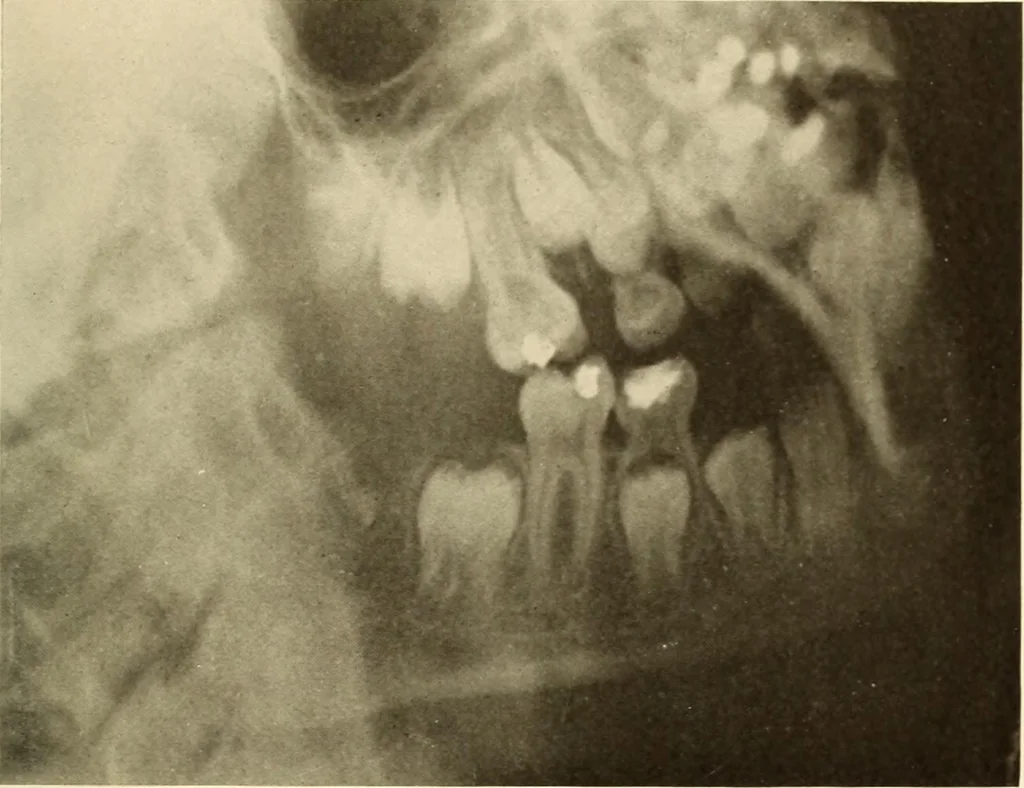
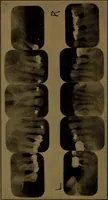
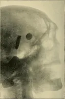
, while the roots of the first permanent molarare completed. Note that the third molars have started to becomecalcified. Figure 16. Patient: G. S., girl twelve years and eight months old. Roentgen Examination: All the temporary teeth have been shed andthe permanent ones have taken their places. The… … Figure 15.