Oral Roentgenology : a Roentgen study of the anatomy and pathology of the oral cavity (1917)
Thoma, Kurt H. (Kurt Hermann), 1883- · Boston, Mass. : Ritter & Company · leaf 44
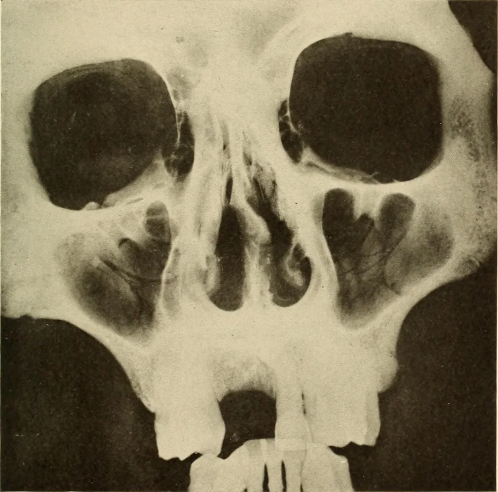
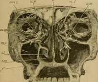
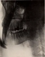
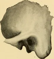
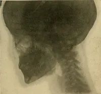
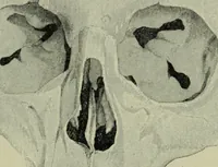
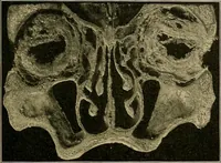
Figure 20. … Figure 40 ORAL ROENTGENOLOGY Roentgenology of the Maxillary Sinuses Figure 22.Patient: Mr. W. V. Roentgen Examination: Shows both of the maxillary sinuses, as wellas the ethmoidal and frontal sinuses in normal condition. ROEXTGEXOGRAPHIC STUDY OF THE XORMAL ORAL TISSUES 41