Oral Roentgenology : a Roentgen study of the anatomy and pathology of the oral cavity (1917)
Thoma, Kurt H. (Kurt Hermann), 1883- · Boston, Mass. : Ritter & Company · leaf 48
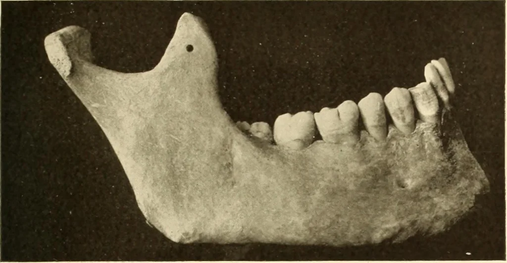
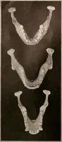
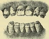
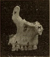
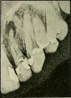
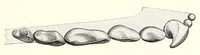
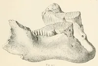
graph: Shows inner structure of bone. The stratum durumlining the alveolar sockets is especially well shown on the two biscupids.The mandibular canal is partly exposed. Figure 25. Specimen: From dry skull shown in Figures 23 and 24. Roentgen Examination: Shows also the inner make-up of the bone.The… … Figure 23.