Oral Roentgenology : a Roentgen study of the anatomy and pathology of the oral cavity (1917)
Thoma, Kurt H. (Kurt Hermann), 1883- · Boston, Mass. : Ritter & Company · leaf 48
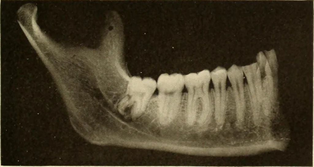
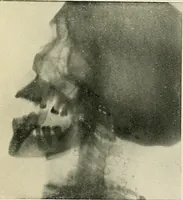
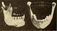
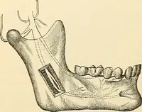
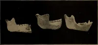
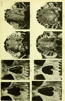
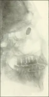
Figure 24. … 44 ORAL ROENTGENOLOGY Roentgenology of the Mandible Figure 26. Specimens: Cross sections of mandible. Photograph: Shows on left a cross section through bicuspid region.Note the thick cortical bone and cancellous inner part; also the mandib-ular canal and mental foramen. On the right a cross section…