Oral Roentgenology : a Roentgen study of the anatomy and pathology of the oral cavity (1917)
Thoma, Kurt H. (Kurt Hermann), 1883- · Boston, Mass. : Ritter & Company · leaf 52
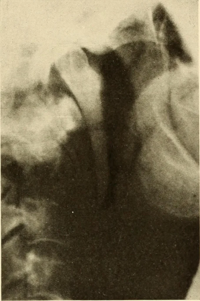
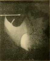
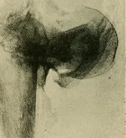
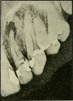
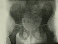
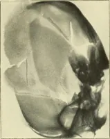
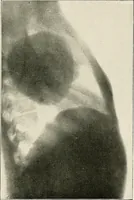
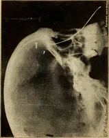
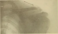
Figure 27. 46 ORAL ROENTGENOLOGY Roentgenology of the Mandible Figure 28.Patient: Mrs. N. Roentgen Examination: Shows normal mandibular joint, coronoidprocess and mandibular notch. Figure 29.Specimen: From dry skull. Roentgen Examination: Shows mandibular canal and distributionto the teeth.… … Figure 28.