Oral Roentgenology : a Roentgen study of the anatomy and pathology of the oral cavity (1917)
Thoma, Kurt H. (Kurt Hermann), 1883- · Boston, Mass. : Ritter & Company · leaf 52
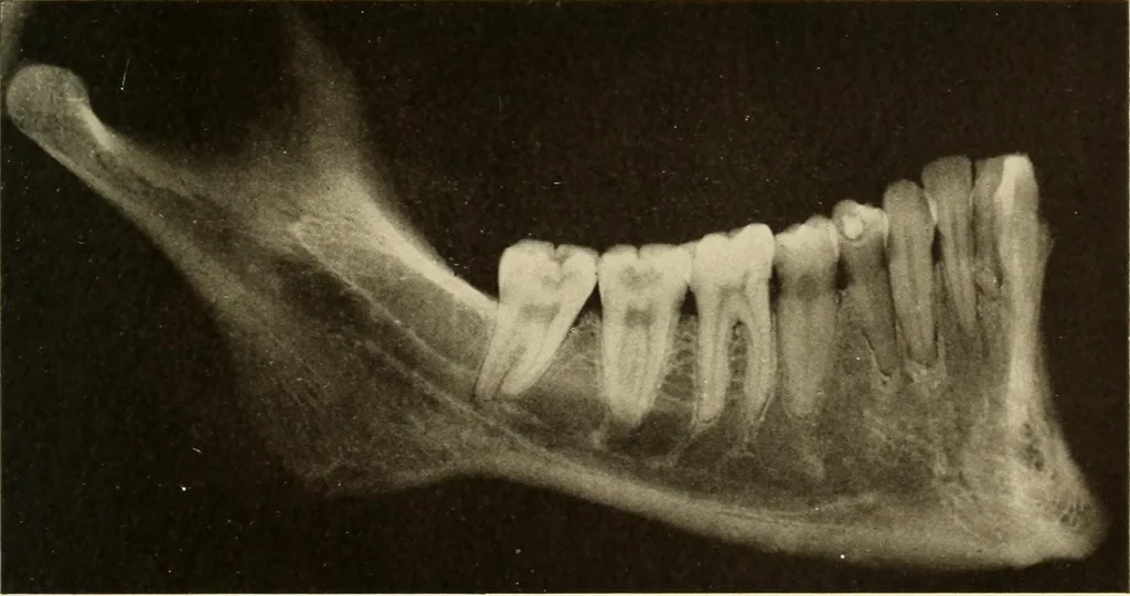
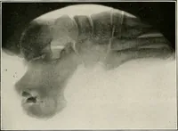
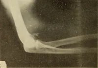
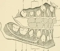
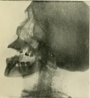
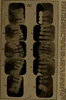
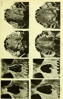
Figure 28. … Figure 29. 48 ORAL ROENTGENOLOGY Roentgenology of Normal Teeth Figure 30. Patient: Miss J., age 11. Roentgen Examination: Shows the roots of the first molar finished,roots of the second bicuspid and second molar not yet completed andthe foramen wide open. Note size of the pulp canals. Figure…