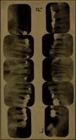
Oral Roentgenology : a Roentgen study of the anatomy and pathology of the oral cavity (1917)
Thoma, Kurt H. (Kurt Hermann), 1883- · Boston, Mass. : Ritter & Company · leaf 68
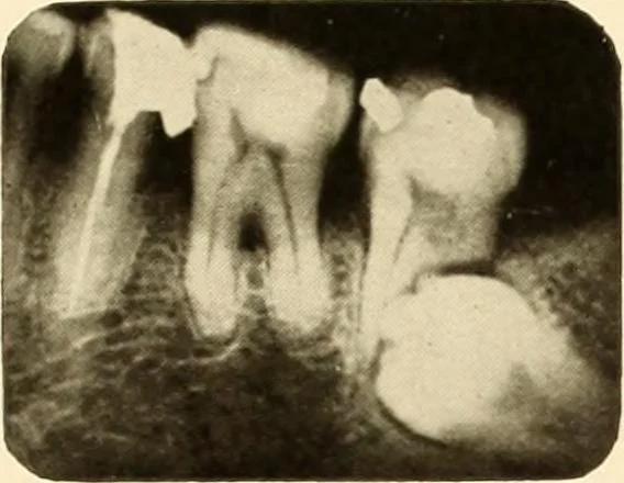
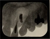
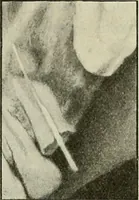
o be extracted.The distal root of the second molar had been completely absorbed andthe distal surface of the mesial root also showed evidence of absorption. Result of Operation: Patient improved a great deal, but was notentirely relieved of headaches. Figure 51. Patient: Dr. E. S. W. Roentgen… … Figure 50.