Oral Roentgenology : a Roentgen study of the anatomy and pathology of the oral cavity (1917)
Thoma, Kurt H. (Kurt Hermann), 1883- · Boston, Mass. : Ritter & Company · leaf 72
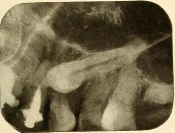
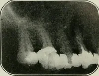
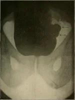
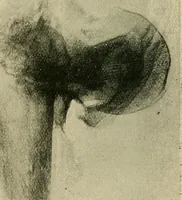
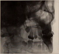
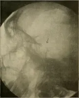
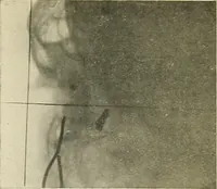
th. The bonewas necrotic and the teeth decayed at the crown. Figure 61. Patient: Mrs. J. C. History: Had pain and swelling in cuspid region of lower jaw. PusAvas discharged from a sinus. Roentgen Examination: Shows unerupted cuspid. Radiolucent areaaround crown of tooth indicates abscess condition…