Oral Roentgenology : a Roentgen study of the anatomy and pathology of the oral cavity (1917)
Thoma, Kurt H. (Kurt Hermann), 1883- · Boston, Mass. : Ritter & Company · leaf 76
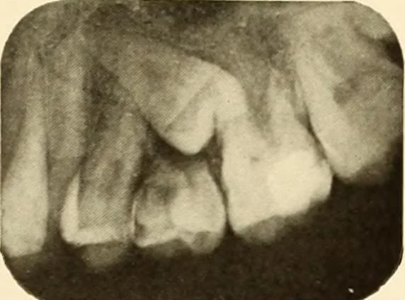
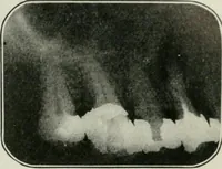
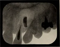
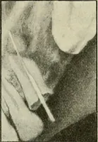
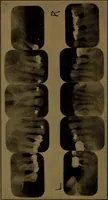
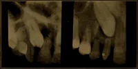
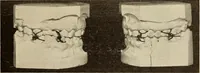
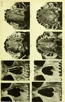
ion: Shows that the unerupted permanent bicuspidis growing in the wrong direction and is impacted. The temporarymolar, which has been retained, shows absorption of the roots. Figure 70. Patient: Mrs. B. Roentgen Examination: Shows that the permanent cuspid is un-erupted and impacted and the… … Figure 68. Figure 69.