Oral Roentgenology : a Roentgen study of the anatomy and pathology of the oral cavity (1917)
Thoma, Kurt H. (Kurt Hermann), 1883- · Boston, Mass. : Ritter & Company · leaf 78
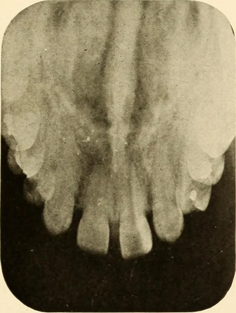
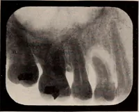
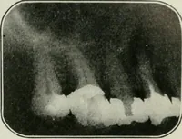
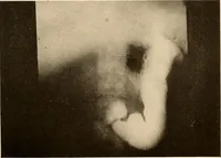
y and permanent lat-eral incisors missing on left side. The picture shows the permanentcentral incisor, the temporary cuspid root, partly absorbed and thepermanent cuspid about two-thirds formed. Figure 73.Patient: Mrs. W. Roentgen Examination: Shows unerupted third molar and rudimen-tary fourth… … Figure 71.