Oral Roentgenology : a Roentgen study of the anatomy and pathology of the oral cavity (1917)
Thoma, Kurt H. (Kurt Hermann), 1883- · Boston, Mass. : Ritter & Company · leaf 88
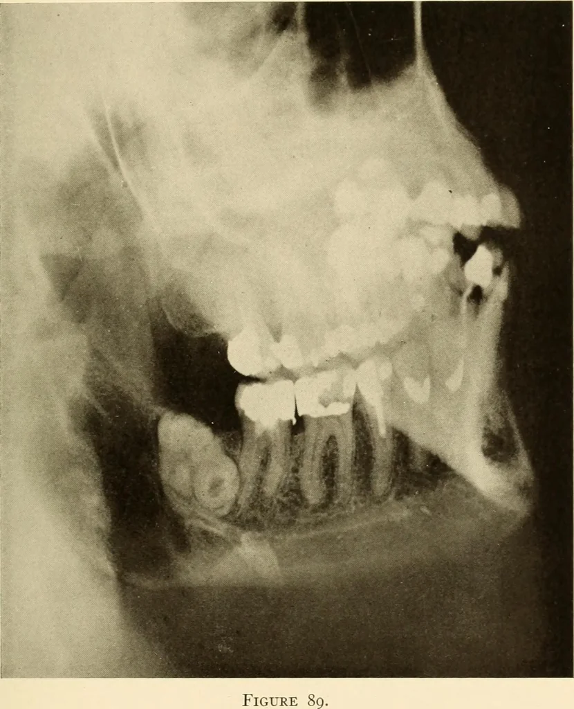
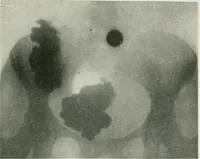
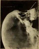
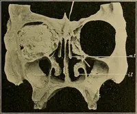
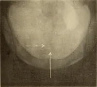
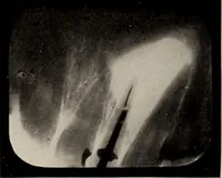
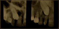
^ ^ V ^ Figure 87. Figure 82 ORAL ROENTGENOLOGY Roentgenology of Odontomata Figure 89. Patient: Mrs. E. B. Roentgen Examination: Examination for infectious foci revealed odon-toma. Operative Findings: The odontoma was made up of two lower molarsgrown together, one in upright and one in transverse… … Figure 84 ORAL ROENTGENOLOGY Roentgenology of Odontomata Figure 90. Patient: Mrs. I. A. V. History: Patient had frequent attacks of severe neuralgia. Roentgen Examination: Shows lower unerupted and impacted wisdomtooth. The wisdom tooth in the upper jaw is also unerupted and hasan extension of…