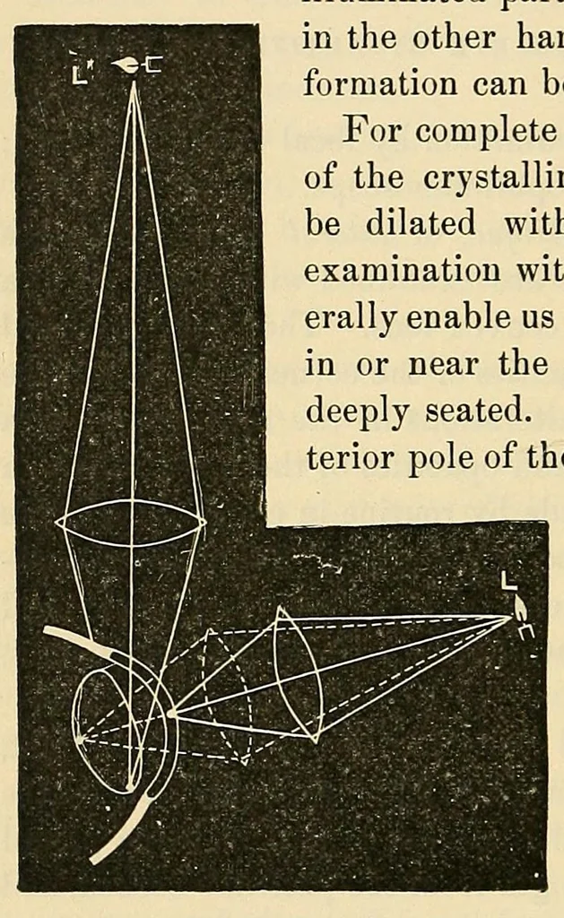
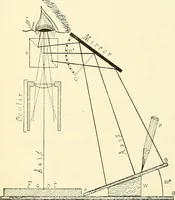
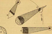
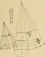
64 FOCAL ILLUMINATION each side, we can examine all parts of the corneal surface, of the iris, of the pupillary area (i. e., the anterior capsule of the lens), and of the lens-substance. If the light be thrown at a very acute angle on the cornea or lens, opaci- ties are much more visible than if it… … and anterior layers of Focal illumination. the lens appear whit- ish, deep opacities in the lens, especially in old people, look yellowish, by focal light. Tumors, large opacities in the vitreous, and retinal detachments may be seen by this method if they lie close behind the lens. Minute foreign…
Eye · Color blindness
Internet Archive · Yale University, Cushing/Whitney Medical Library · Public domain (published before 1931). Free to use for anything.