A text-book of diseases of the nose and throat (1904)
Kyle, David Braden, 1863-1916 · Philadelphia ; New York ; London : W.B. Saunders · leaf 304
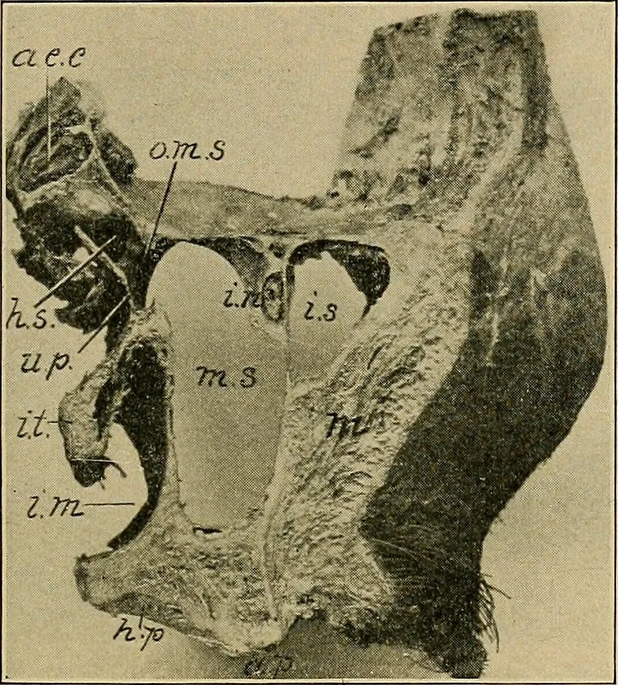
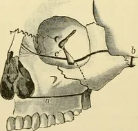
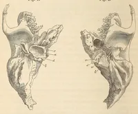
r maxillary bone (Fig. 2), are the largest of the connectednasal structures. Anatomically, a brief study of each cavity, to thearticle on which the reader is referred, shows many peculiarities 278 DISEASES OF THE ANTERIOR NASAL CAVITIES. 279 favorable to the origin of morbid processes. Fig. 95… … Fig. 95.—Anterior wall of antrum removed (after Cryer): a.e.c, anterior ethmoidalcells; h.s., hiatus semilunaris; u.p., uncinate process; m.s., maxillary sinus; i.t., inferiorturbinate; i.m., inferior meatus; o.m.s., opening into maxillary sinus; i.n., infra-orbitalnerve; m., muscles of face; h.p.,…