Illustrations of disease with the microscope. clinical investigations, aided by the microscope, and by chemical reagents, with microscopical observations of pathological specimens, medical and surgical, obtained in Charleston, S.C. : a contribution intended to disclose the minute history of the diseases prevailing in this latitude, and to assist future students (1861)
Porcher, Francis Peyre, 1825-1895, Billings, John S. (John Shaw), 1838-1913 · Charleston, S.C., C.S.A. : South Carolina Medical Association · leaf 102
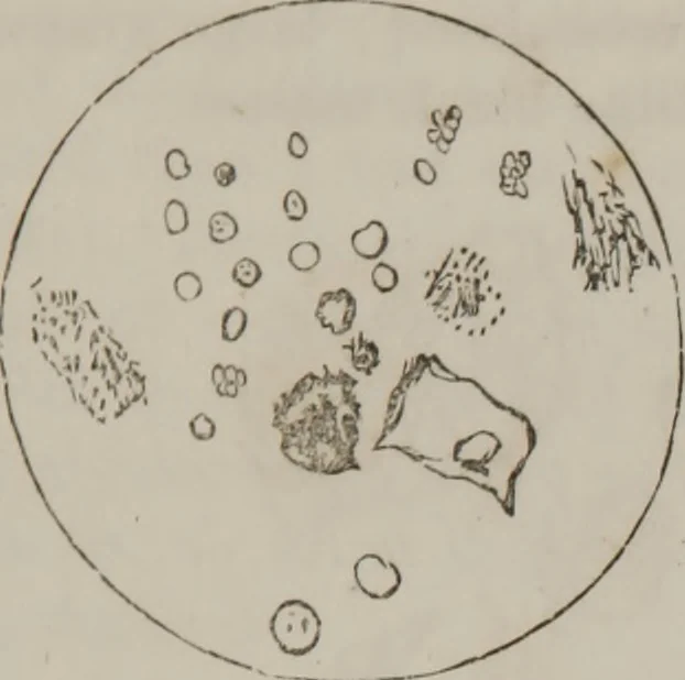
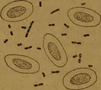
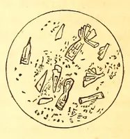
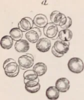
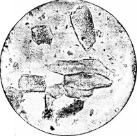
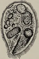
Fig. 2. WITH THE MICROSCOPE. 95 … Another specimen examined 26tk.Patient is recovering as it were by a miracle. Urine now free ofblood, clear amber color, }-et someslirunk-np blood corpuscles undermicroscope, see Fig. 3. Xo crys-tals ; epithel. scales; not muchalbuminous granular matter. Pig. 3.—Disks at right of figure small,…
Microscopy
Internet Archive · Medical Heritage Library · Public Domain Mark 1.0. Free to use for anything.