Illustrations of disease with the microscope. clinical investigations, aided by the microscope, and by chemical reagents, with microscopical observations of pathological specimens, medical and surgical, obtained in Charleston, S.C. : a contribution intended to disclose the minute history of the diseases prevailing in this latitude, and to assist future students
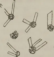
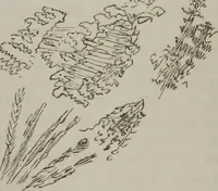
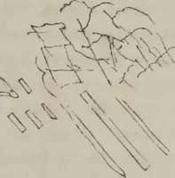
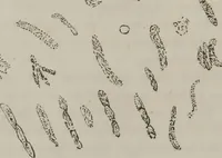
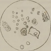
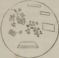
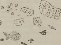
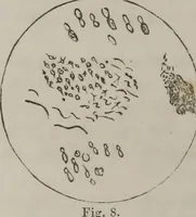
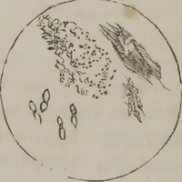
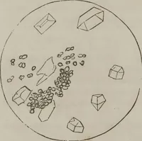
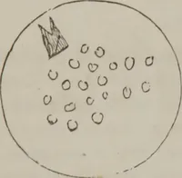
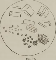
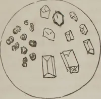
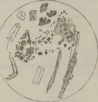
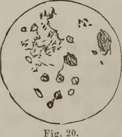
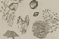
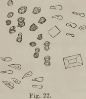
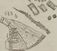
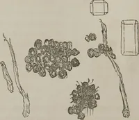
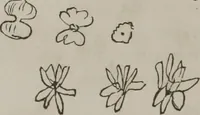
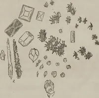
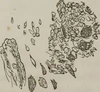
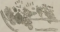
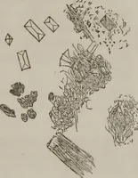
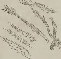
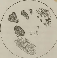
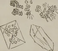
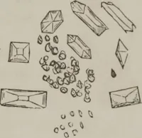
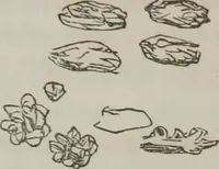
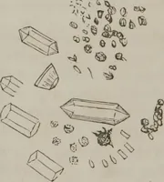
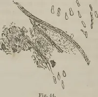
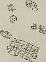
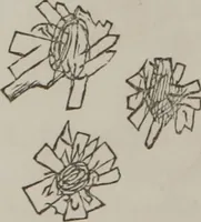
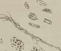
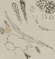
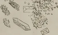
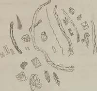
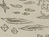
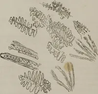
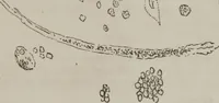
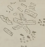
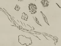
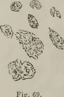
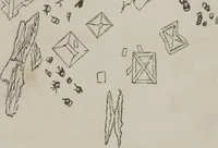
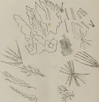
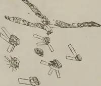
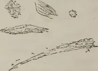
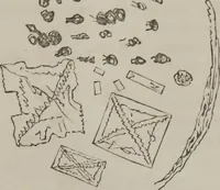
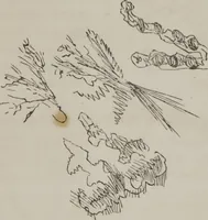
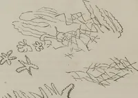
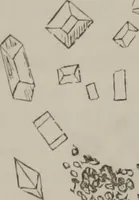
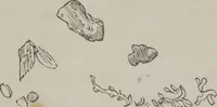
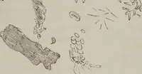
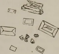
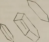
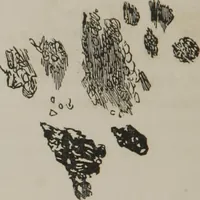
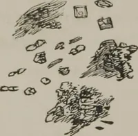
A clinical microscopy atlas by Charleston physician Francis Peyre Porcher, issued in 1861 by the South Carolina Medical Association for local doctors and medical students recording pathological specimens seen under the lens. The plates are sparse pen drawings scattered across bare paper: faceted crystals, lumpy cell clusters, stringy casts and fibre bundles, useful as isolated organic marks, scatter patterns and small abstract specimen silhouettes.