Illustrations of disease with the microscope. clinical investigations, aided by the microscope, and by chemical reagents, with microscopical observations of pathological specimens, medical and surgical, obtained in Charleston, S.C. : a contribution intended to disclose the minute history of the diseases prevailing in this latitude, and to assist future students (1861)
Porcher, Francis Peyre, 1825-1895, Billings, John S. (John Shaw), 1838-1913 · Charleston, S.C., C.S.A. : South Carolina Medical Association · leaf 102
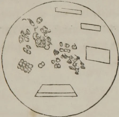
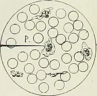
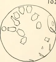
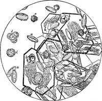
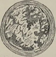
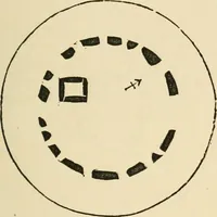
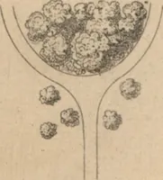
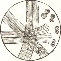
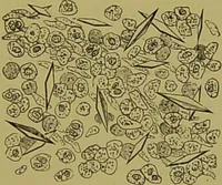
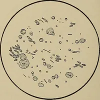
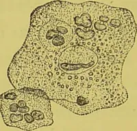
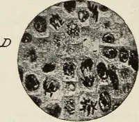
Fig. I. Fig. 5. Case 2d.—August, 1856. Mathews: Specimen sent fromRoper Hospital; sick three weeks.First case—resembling, if not clearlyYellow Fever. Patient taking inius.serpentaria, chlorate of potash, andquinine. Urate of soda and amnion., magnes.Phosphates under microscope. Fig. G. One… … round white bodies; 96 ILLUSTRATIONS OF DISEASE blood corpuscles, small, wrinkled, crenulated : large granu-lar masses, no crystals, non-crystalline black masses. V
Microscopy
Internet Archive · Medical Heritage Library · Public Domain Mark 1.0. Free to use for anything.