Illustrations of disease with the microscope. clinical investigations, aided by the microscope, and by chemical reagents, with microscopical observations of pathological specimens, medical and surgical, obtained in Charleston, S.C. : a contribution intended to disclose the minute history of the diseases prevailing in this latitude, and to assist future students (1861)
Porcher, Francis Peyre, 1825-1895, Billings, John S. (John Shaw), 1838-1913 · Charleston, S.C., C.S.A. : South Carolina Medical Association · leaf 108
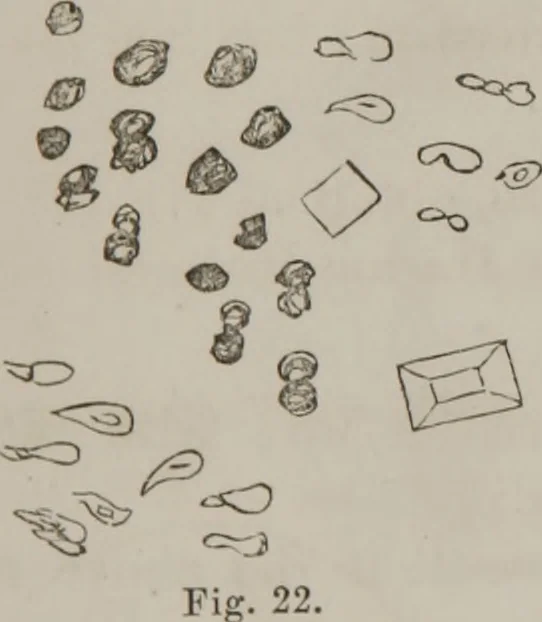
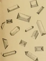
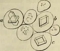
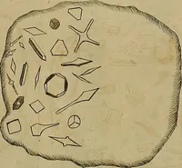
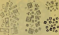
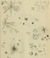
ular matter. Fig. 21. Examined by Reagents With application of heat, albu-men abundant; coagulated and separated by addition ofnitric acid ; some fermentation ; no purpurine ; slight fer-mentation when soda is added. Case 1th.—Specimen sent on 15th, examined same day.Peterson: Roper Hospital;… … Lower strata: Presentedaccompanying microscopicalappearances—objects in Figs.23 and 24 unusually large,visible to naked eye : Fig.23, triple phosphates andurate of soda.
Microscopy
Internet Archive · Medical Heritage Library · Public Domain Mark 1.0. Free to use for anything.