Illustrations of disease with the microscope. clinical investigations, aided by the microscope, and by chemical reagents, with microscopical observations of pathological specimens, medical and surgical, obtained in Charleston, S.C. : a contribution intended to disclose the minute history of the diseases prevailing in this latitude, and to assist future students (1861)
Porcher, Francis Peyre, 1825-1895, Billings, John S. (John Shaw), 1838-1913 · Charleston, S.C., C.S.A. : South Carolina Medical Association · leaf 111
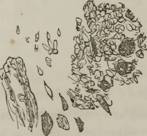
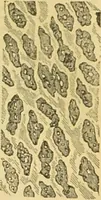
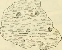
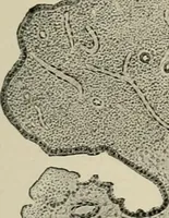
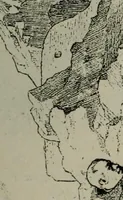
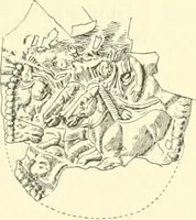
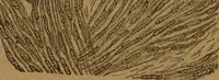
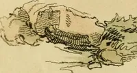
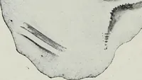
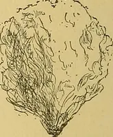
Fi<j. 28. Action of Reagents: But slightly affected by heat; bynitric acid, darkened more than that obtained on 17th, rela-tively (purpurine) ; no particular increase ol sediment. 104 ILLUSTRATIONS OF DISEASE Bile scarcely appreciable by tesl ; alkaline.Vegetable growths even more distinct after… … The upper surface presented, un-der microscope, the appearancesdepicted in Fig. 29: Light yellow,somewhat granular matter, withdark, round bodies ; blood red—thicker; confervoid growths. Fig. 29. Lower strata, represented by Fig. 30 : Dark crystals,round, like urate of soda ? It is difficult to…
Microscopy
Internet Archive · Medical Heritage Library · Public Domain Mark 1.0. Free to use for anything.