Illustrations of disease with the microscope. clinical investigations, aided by the microscope, and by chemical reagents, with microscopical observations of pathological specimens, medical and surgical, obtained in Charleston, S.C. : a contribution intended to disclose the minute history of the diseases prevailing in this latitude, and to assist future students (1861)
Porcher, Francis Peyre, 1825-1895, Billings, John S. (John Shaw), 1838-1913 · Charleston, S.C., C.S.A. : South Carolina Medical Association · leaf 111
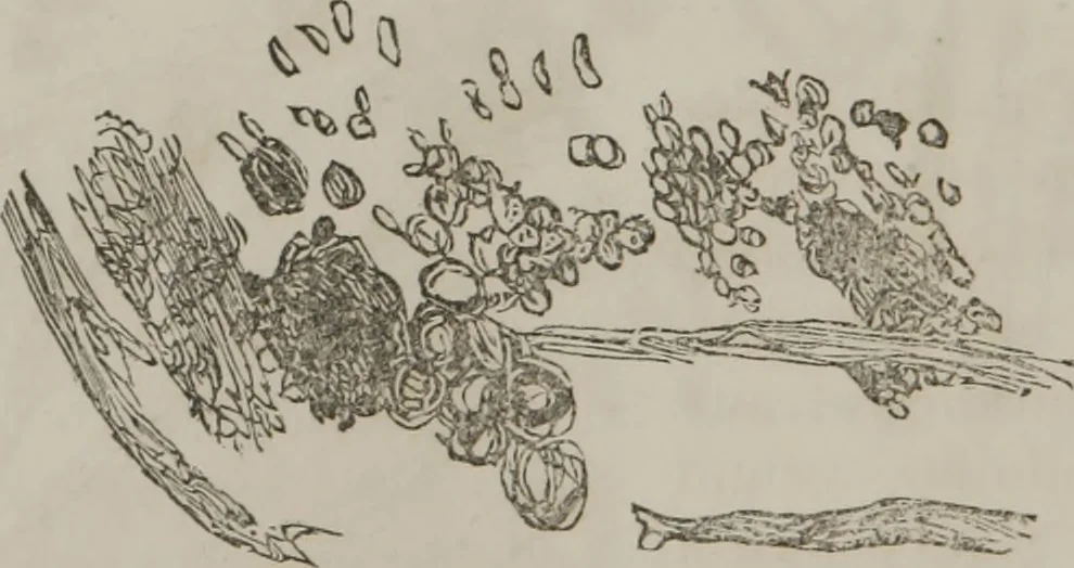
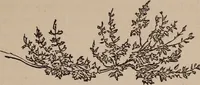
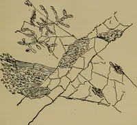
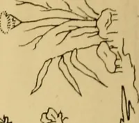
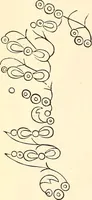
n Fig. 29: Light yellow,somewhat granular matter, withdark, round bodies ; blood red—thicker; confervoid growths. Fig. 29. Lower strata, represented by Fig. 30 : Dark crystals,round, like urate of soda ? It is difficult to understandhow such bodies, which are plainly urate of soda, shouldhave… … 30. Specimen from same, collected 17th, examined 17th : Clearamber color; mucus-looking particles floating in it. Action of Reagents: Not thickened or turbid by heat, but U IT 11 THE .MICROSCOPE. 105 very much darkened by nitric acid, showing purpurine,with effervescence. Case Hth.— Specimen…
Microscopy
Internet Archive · Medical Heritage Library · Public Domain Mark 1.0. Free to use for anything.