Illustrations of disease with the microscope. clinical investigations, aided by the microscope, and by chemical reagents, with microscopical observations of pathological specimens, medical and surgical, obtained in Charleston, S.C. : a contribution intended to disclose the minute history of the diseases prevailing in this latitude, and to assist future students (1861)
Porcher, Francis Peyre, 1825-1895, Billings, John S. (John Shaw), 1838-1913 · Charleston, S.C., C.S.A. : South Carolina Medical Association · leaf 112
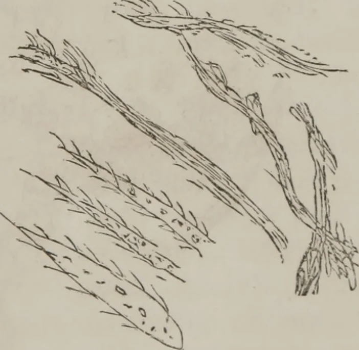
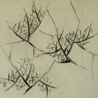
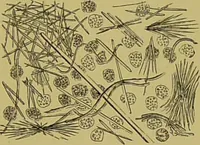
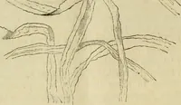
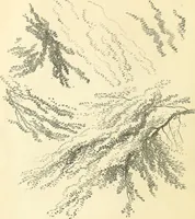
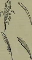
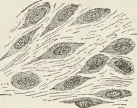
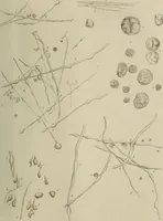
Fig. 32. These (Fig. 33) proba-bly foreign substances :hairs, bits of bed clothing,etc.; some have strange-ly the appearance of ani-mals : Polonius wouldhave said, very like afish. Fig. 31. Lower strata: Pale, chalkyand flaky sediment; ani-moniacal odor; against thelight, urine in vial has… … Fiji. 33. Action of Beagents: Heat did not coagulate it, or make itmore turbid; with nitric acid, effervesced so actively that 10>3 ILLUSTRATIONS OF DISEASE nearly all in the test tube escaped: did not coagulate ; itbecame slightly darker, then cleared up. There was seenafterward some separated…
Microscopy
Internet Archive · Medical Heritage Library · Public Domain Mark 1.0. Free to use for anything.