Illustrations of disease with the microscope. clinical investigations, aided by the microscope, and by chemical reagents, with microscopical observations of pathological specimens, medical and surgical, obtained in Charleston, S.C. : a contribution intended to disclose the minute history of the diseases prevailing in this latitude, and to assist future students (1861)
Porcher, Francis Peyre, 1825-1895, Billings, John S. (John Shaw), 1838-1913 · Charleston, S.C., C.S.A. : South Carolina Medical Association · leaf 114
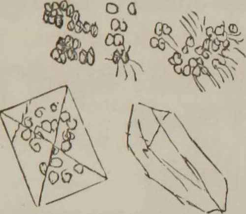
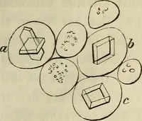
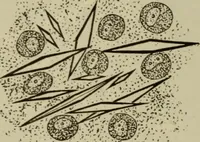
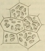
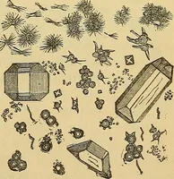
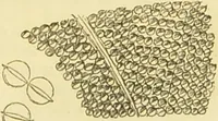
Fig. .35* WITH TJIE MICROSCOPE. 107 Case 11th.—Specimen obtained 17th, examined 17th Sept.,1856. KeUigan: Roper Hospital; Yellow Fever—not dis-tinctly marked. Specimen exhibited dark froth, as iftinged by bile. Action of Reagents: With heat, thickening; very albumi-nous; nitric acid separated the… … Same examined 18th. Upper sur-face under microscope, Fig. 36. Fig. 36. Lower stratum : Yellow, flocu-lent deposit abundant; urate ofsoda and triple phosphates. Thesmall, circular disks in lowerpart of figure were clear and pel-lucid, resembling organic glob-ules.
Microscopy
Internet Archive · Medical Heritage Library · Public Domain Mark 1.0. Free to use for anything.