Illustrations of disease with the microscope. clinical investigations, aided by the microscope, and by chemical reagents, with microscopical observations of pathological specimens, medical and surgical, obtained in Charleston, S.C. : a contribution intended to disclose the minute history of the diseases prevailing in this latitude, and to assist future students (1861)
Porcher, Francis Peyre, 1825-1895, Billings, John S. (John Shaw), 1838-1913 · Charleston, S.C., C.S.A. : South Carolina Medical Association · leaf 114
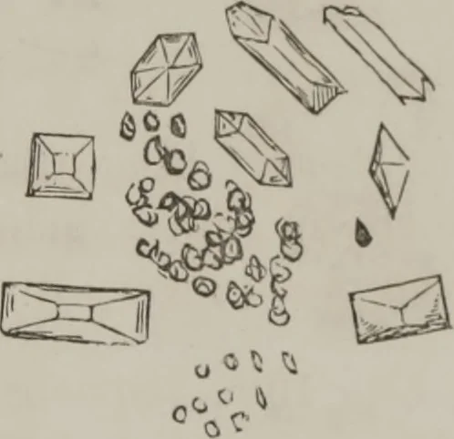
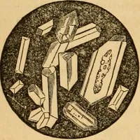
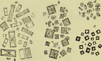
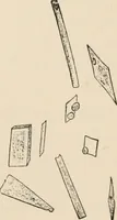
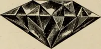
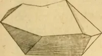
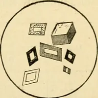
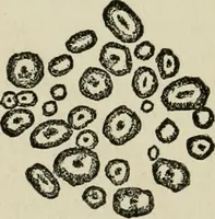
Same examined 18th. Upper sur-face under microscope, Fig. 36. Fig. 36. Lower stratum : Yellow, flocu-lent deposit abundant; urate ofsoda and triple phosphates. Thesmall, circular disks in lowerpart of figure were clear and pel-lucid, resembling organic glob-ules. … Fig. 37. Action of Reagents: Examined with heat: did not thicken,but became a little clearer, though leaving shreds ; frotheda great deal ; hue greenish ; when nitric arid was added,nearly all boiled over; did not turn near so dark as on thedav before. A great deal of sedimentary matter…
Microscopy
Internet Archive · Medical Heritage Library · Public Domain Mark 1.0. Free to use for anything.