Illustrations of disease with the microscope. clinical investigations, aided by the microscope, and by chemical reagents, with microscopical observations of pathological specimens, medical and surgical, obtained in Charleston, S.C. : a contribution intended to disclose the minute history of the diseases prevailing in this latitude, and to assist future students (1861)
Porcher, Francis Peyre, 1825-1895, Billings, John S. (John Shaw), 1838-1913 · Charleston, S.C., C.S.A. : South Carolina Medical Association · leaf 115
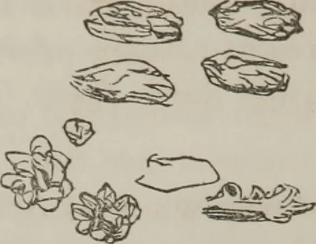
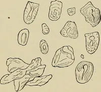
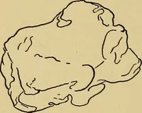
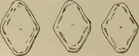
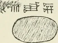
t turn near so dark as on thedav before. A great deal of sedimentary matter separated,showing that albumen is exhibited on the second day.Iritie hue, showing bile, on white plate : alkaline reaction. Note (made subsequently).— In another specimen ofKelligans ? urine found in a test tube (to which… … FiK. 38. Shcmpfcls case, further on, withthe addition of crystals of dark,red hue floating in the tube, manyof them regular; nitric acid dis-solves them, with evolution ofbubbles. The crystals, Fig. 38,are undoubtedly those of uric acid. Case 12th.—Specimen obtained 18th, evening, examined19th.…
Microscopy
Internet Archive · Medical Heritage Library · Public Domain Mark 1.0. Free to use for anything.