Illustrations of disease with the microscope. clinical investigations, aided by the microscope, and by chemical reagents, with microscopical observations of pathological specimens, medical and surgical, obtained in Charleston, S.C. : a contribution intended to disclose the minute history of the diseases prevailing in this latitude, and to assist future students (1861)
Porcher, Francis Peyre, 1825-1895, Billings, John S. (John Shaw), 1838-1913 · Charleston, S.C., C.S.A. : South Carolina Medical Association · leaf 116
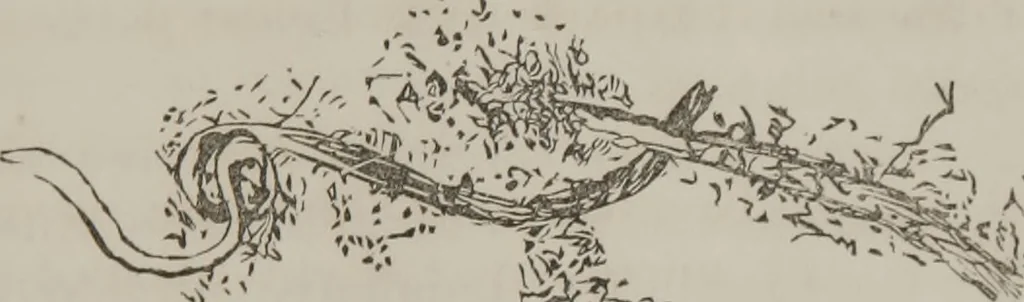
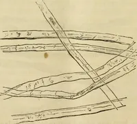
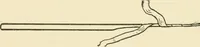
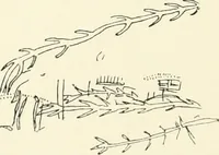
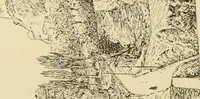
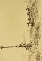
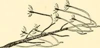
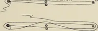
. 18th, evening, ex-amined 19th. Shempfel: Roper Hospital. WITH THK .MICROSCOPE. 109 Appearance of specimen : Reddish colored, dark frothon surface, stained with bile; red flakes at bottom of vial. Action of Reagents: Heat very much thickened the entiremass; not so dark; nitric acid did not… … Fig. 42. Red, albuminous-looking granular matter; the flakes in-vesting the tube, which is of vermilion color, look undernnked eye like clotted blood, as in Fig. 44, also. Nocrystals or blood corpuscles. Action of Reagents.—Examined on 20th for bile ; no col-oration on white porcelain; acid…
Microscopy
Internet Archive · Medical Heritage Library · Public Domain Mark 1.0. Free to use for anything.