Illustrations of disease with the microscope. clinical investigations, aided by the microscope, and by chemical reagents, with microscopical observations of pathological specimens, medical and surgical, obtained in Charleston, S.C. : a contribution intended to disclose the minute history of the diseases prevailing in this latitude, and to assist future students (1861)
Porcher, Francis Peyre, 1825-1895, Billings, John S. (John Shaw), 1838-1913 · Charleston, S.C., C.S.A. : South Carolina Medical Association · leaf 117
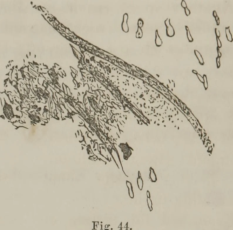
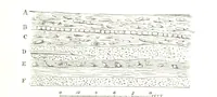
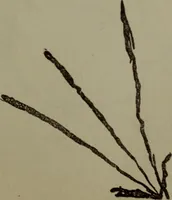
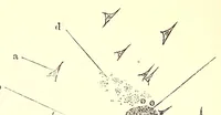
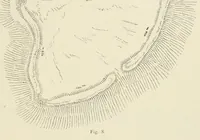
s.—Examined on 20th for bile ; no col-oration on white porcelain; acid reaction; same examined20th, with microscope: §8tt4 <5 W*k ^ o 0,(5 ,^ Surface: Vegetable growths,while, transparent, round disksand epithel. scales. Fig. 13. 110 ILUSTKATIONS OF DISEASE Lover si,-alu (Fig. 44):Dark red,… … Action of Reagents.—Boiled with liquor potassae, etc.;sugar decidedly indicated. Note.—The specimen collected the 18th,and then mixed with nitric acid in test tube.Examined 21st, the albuminous-looking resi-due I find now to be fine, brownish andgranular; no crystals or other substance.See Fig. 45.…
Microscopy
Internet Archive · Medical Heritage Library · Public Domain Mark 1.0. Free to use for anything.