Illustrations of disease with the microscope. clinical investigations, aided by the microscope, and by chemical reagents, with microscopical observations of pathological specimens, medical and surgical, obtained in Charleston, S.C. : a contribution intended to disclose the minute history of the diseases prevailing in this latitude, and to assist future students (1861)
Porcher, Francis Peyre, 1825-1895, Billings, John S. (John Shaw), 1838-1913 · Charleston, S.C., C.S.A. : South Carolina Medical Association · leaf 120
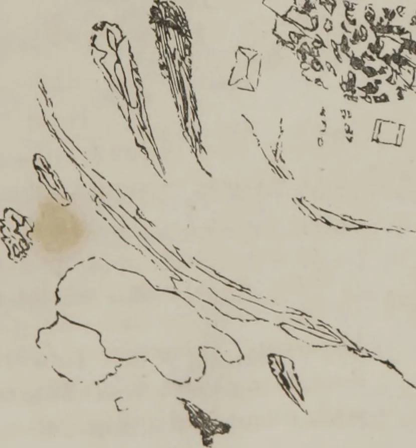
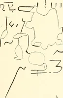
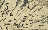
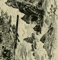
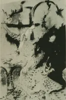
o Fig. 53. Lower strata white, chalky, and filled with urate of sod;.and triple phosphates; wedge-shaped bodies, films of hair.cloth, etc., with large epithel. scale. [Artist has not don<justice to my drawing.] … 114 ILLUSTRATIONS OF DISEASE In a note appended to tins ease, I find written: amongspecimens of urine on my table, collected during the pasteight days ; none have confervoid growths. Case 16///.—Specimen obtained 22d, examined 24th Sept.,1856. Sent from Roper Hospital. Furlong: Renal excre-tion…
Microscopy
Internet Archive · Medical Heritage Library · Public Domain Mark 1.0. Free to use for anything.