Illustrations of disease with the microscope. clinical investigations, aided by the microscope, and by chemical reagents, with microscopical observations of pathological specimens, medical and surgical, obtained in Charleston, S.C. : a contribution intended to disclose the minute history of the diseases prevailing in this latitude, and to assist future students (1861)
Porcher, Francis Peyre, 1825-1895, Billings, John S. (John Shaw), 1838-1913 · Charleston, S.C., C.S.A. : South Carolina Medical Association · leaf 122
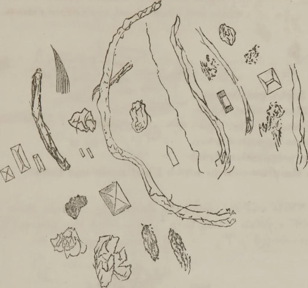
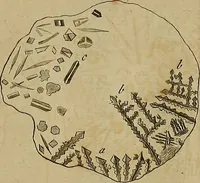
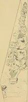
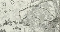
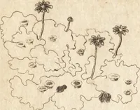
Fie. Same specimen examined 25th. Action oflleagents: Strong alkaline reaction ; thick, lightish-yellow color; foamed overand thickened by beat; with nitric acid, nearly all wasted ;thickened, but with no sediment like albumen ; no suear. Specimen of 26th ? examined 27th. Lower stratum, seeFig.… … Fig. 58. Specimen of renal discharge in Furlongs case—thoughkepi for two months, still contained vibriones in activemotion. Examined Febsize—quite as sm57. >. 19th: Not increased in A1 I /J/all : very active. See Fig- \ (rj^ \ 1 Jf& Fig. 57. Case Vlth.—Specimen sent Sept. 18th, examined…
Microscopy
Internet Archive · Medical Heritage Library · Public Domain Mark 1.0. Free to use for anything.