Illustrations of disease with the microscope. clinical investigations, aided by the microscope, and by chemical reagents, with microscopical observations of pathological specimens, medical and surgical, obtained in Charleston, S.C. : a contribution intended to disclose the minute history of the diseases prevailing in this latitude, and to assist future students (1861)
Porcher, Francis Peyre, 1825-1895, Billings, John S. (John Shaw), 1838-1913 · Charleston, S.C., C.S.A. : South Carolina Medical Association · leaf 123
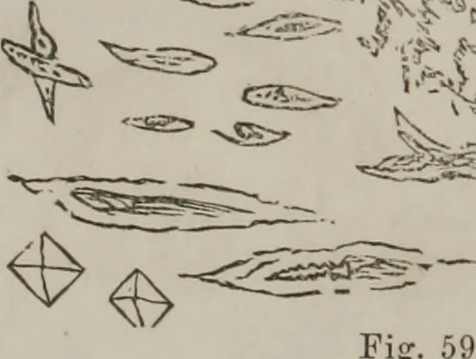
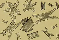
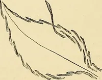
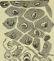
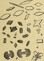
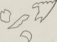
Upper surface: Small, rounded bodwhite and clear; hard,crystalline masses. ies — Fig. 58. Light, purple, crys-talline bodies, centreprismatic, Fig. 59.—These appeared whenthe preparation dried V - … ESS ^P^fV^ on the glass—forms which I had never met before. Nitric acid converted the above, Fig. 59, into these den-dritic crystals (stag-horn), dissolving the unusual formsrepresented in Fig. 59, and lea vino- these, Fig. 60.
Microscopy
Internet Archive · Medical Heritage Library · Public Domain Mark 1.0. Free to use for anything.