Illustrations of disease with the microscope. clinical investigations, aided by the microscope, and by chemical reagents, with microscopical observations of pathological specimens, medical and surgical, obtained in Charleston, S.C. : a contribution intended to disclose the minute history of the diseases prevailing in this latitude, and to assist future students (1861)
Porcher, Francis Peyre, 1825-1895, Billings, John S. (John Shaw), 1838-1913 · Charleston, S.C., C.S.A. : South Carolina Medical Association · leaf 128
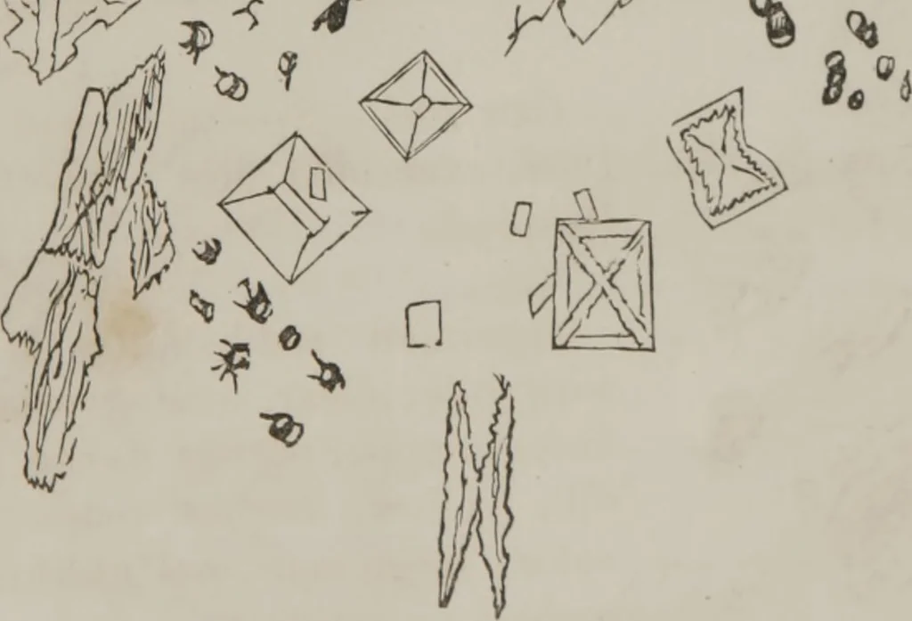
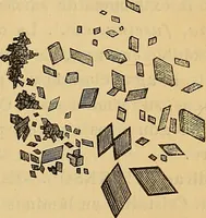
only atrace. Case 2\st.—Obtained 22d Sept., 1856, examined 24th.Conner or Connel: Roper Hospital. Renal excretion yellow,colored by bile; froth also tinged yellow. Action of Reagents: The chalky deposit, when heated,almost wasted by frothing away; no deposit with heat. A sediment in test tube after… … Fia 122 [LLUSTRATIONS <>!• DISB \.SE Specimen of 26th. Paler, reddish yellow. Treated by Reagents: Albuminous by heat; the albumenseparated by nitric acid, without any froth from eitheragent; some discoloration (like purpurine); the color turnsto a very rich, dark yellow: no greenish…
Microscopy
Internet Archive · Medical Heritage Library · Public Domain Mark 1.0. Free to use for anything.