Illustrations of disease with the microscope. clinical investigations, aided by the microscope, and by chemical reagents, with microscopical observations of pathological specimens, medical and surgical, obtained in Charleston, S.C. : a contribution intended to disclose the minute history of the diseases prevailing in this latitude, and to assist future students (1861)
Porcher, Francis Peyre, 1825-1895, Billings, John S. (John Shaw), 1838-1913 · Charleston, S.C., C.S.A. : South Carolina Medical Association · leaf 134
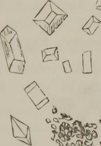
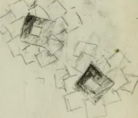
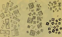
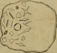
ow and sedimentary, 1ml cer-tainly very great improvement, judging from the renalexcretion ; reaction neutral; sugar none; no sediment withliquor potassse. Case 23d.—Specimen obtained 22d, examined 24th Sept.,1856. Tobin: Roper Hospital. Pale colored, abundant,white, chalky sediment. WfTIf THE… … s?>^ a fl.f* Sir Lower stratum: White, chalkydeposit, composed of uratesand phosphates, Fig. 82. <^v> Fig. 82. Case 24//;.—Examined 11th, collected 10th Sept., 1856.Fitzgerald: Roper Hospital. Dark colored, slightly crys-talline masses present, some epithelial scales; circularbodies larger…
Microscopy
Internet Archive · Medical Heritage Library · Public Domain Mark 1.0. Free to use for anything.